The study of terrestrial fossils in ancient rocks: a crucial approach to identify potential signs of life on Mars

The NASA Perseverance rover is actively exploring Jezero Crater, analyzing igneous and sedimentary rocks from the crater floor and delta deposits. The rock samples that will be returned by the Mars Sample Return (MSR) mission in the 2030s will be subjected to detailed laboratory studies.

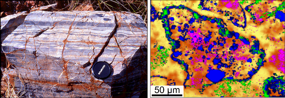
Some samples may contain traces of ancient Martian life, which are challenging to detect due to their morphological simplicity and subtle geochemical expressions. Using volcanic sediments from Kitty’s Gap Chert (Pilbara, Australia) of 3.45 billion years as analogues, researchers detail the steps needed to demonstrate their syngenicity and biogenicity. Various analytical methods, including optical and electron microscopy, Raman spectroscopy, X-ray fluorescence spectroscopy, and mass spectrometry, have been employed at different scales. Sedimentological, petrological, mineralogical, and geochemical analyses document a coastal environment of deposition, consistent with the development of microbial life. Morphological, elemental, and molecular analyses of carbonaceous matter associated with potential fossil remnants reveal enrichment in bioessential trace metals (V, Cr, Fe, Co, etc.) and colocalized aromatic and aliphatic molecules of biological origin. This study illustrates the analytical protocol necessary to optimize the detection of fossil traces of life in Martian rocks.

This work is reported on the CNRS Chimie website

Reference
Multi-Technique Characterization of 3.45 Ga Microfossils on Earth: A Key Approach to Detect Possible Traces of Life in Returned Samples from Mars
Laura Clodoré, Frédéric Foucher, Keyron Hickman-Lewis, Stéphanie Sorieul, Jean Jouve, Matthieu Réfrégiers, Guillaume Collet, Stéphane Petoud, Bernard Gratuze, Frances Westall
Astrobiology 2024
http://doi.org/10.1089/ast.2023.0089

Enzymatic detection in near infrared optical imaging and MRI with a single ligand complexed to different lanthanide ions

The imaging visualization of active enzymes is of primary importance in biology.

In a collaborative effort between the Centre of Molecular Biophysics (CBM) and the Institute of Chemistry of Natural Substances (ICSN) in Gif sur Yvette, CBM researchers have designed Ln3+ complexes that provide enzyme-mediated changes in NIR luminescence, as well as in Chemical Exchange Saturation Transfer (CEST) and classical T1-weighted MRI, depending on the Ln3+ used. They have demonstrated the successful monitoring of b-galactosidase activity over time in NIR luminescence and CEST MR imaging in phantoms containing the Yb-complex, and in T1 MRI when using the Gd-analogue. A further great advantage of their probe design is its high versatility, as there are a large number of enzymatically cleavable groups that could be attached to the same core, thus creating probes for other important enzyme targets.

Reference : Rémy Jouclas, Sophie Laine, Svetlana V. Eliseeva, Jérémie Mandel, Frédéric Szeremeta, Pascal Retailleau, Jiefang He, Jean-Francois Gallard, Agnès Pallier, Célia S. Bonnet, Stéphane Petoud, Philippe Durand, Éva Tóth
Lanthanide-Based Probes for Imaging Detection of Enzyme Activities by NIR Luminescence, T1- and ParaCEST MRI
Angew. Chem. Int. Ed. 2024, https://onlinelibrary.wiley.com/doi/10.1002/anie.202317728

Marie Curie Postdoctoral Fellowships

Rafael Aroso, from the University of Coimbra, Portugal, will join the “Metal Complexes and MRI” team. Its “PorphIRON” project aims to develop paramagnetic complexes based on transition metals, such as iron, for MRI applications. This work is part of the team's efforts to replace gadolinium, currently used in MRI, with more biocompatible and ecologically more sustainable metals.

Ross Ballantine, from Queen's University Belfast in Northern Ireland, will join the "Synthetic Proteins and Bioorthogonal Chemistry" team to work on the « ThioSHowcase » project. This project aims to explore arylthiol-based amino acids for the chemoselective functionalisation of peptides and of proteins,  with applications in protein labeling and selective peptide cross-linking.

New imaging probe enables MRI detection of early-stage breast cancer tumors

Despite significant progress in cancer imaging and treatment over the years; early diagnosis, metastasis detection, and a better understanding of cancer progression remain an unmet clinical need. Molecular imaging can fulfil this need, but requires the design of contrast agents which target specific tumor biomarkers. Netrin-1 is an extracellular protein overexpressed in metastatic breast, and it is implicated in tumor progression, angiogenesis and appearance of metastasis and tumor aggressivity.

Reserachers of the team "Metal complexes and MRI", develop and validated in vivo the first Netrin-1 specific peptide-based multimodal probe.  A structural validation was performed by molecular docking, thanks to a collaboration CBM and Institute of Organic and Analytical Chemistry.

This probe enabled successful triple-negative metastatic breast tumor visualization namely at early development stage (tumor volume 0-50 mm3), by magnetic resonance imaging (MRI). Such peptide-based probes are molecular platforms allowing imaging by MRI or single photon emission tomography. This bimodal approach makes it possible to scan a wide range of target concentrations (nM to μM), enabling in vivo mapping of Netrin-1 in cancer murine models, at different tumor evolution stages.

References:
Clémentine Moreau, Tea Lukačević, Agnès Pallier, Julien Sobilo, Samia Aci-Sèche, Norbert Garnier, Sandra Même, Éva Tóth and Sara LacerdaPeptide-Conjugated MRI Probe Targeted to Netrin-1, a Novel Metastatic Breast Cancer Biomarker
Bioconjugate Chem. https://doi.org/10.1021/acs.bioconjchem.3c00558