Structural insights into the SUMOylation reaction

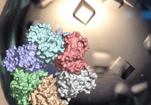
SUMOylation and ubiquitylation are related protein modifications where small proteins (SUMO or ubiquitin) become covalently attached to protein substrates to regulate their function. Both these protein modifications are essential for viability and are strongly implicated in human disease, but SUMOylation remains less studied than ubiquitylation. A key step in both SUMOylation and ubiquitylation reactions is the formation of a reactive thioester molecule in which SUMO or ubiquitin becomes linked to a cysteine residue on proteins called E2. It is from there that SUMO/ubiquitin is transferred onto the final protein substrate. In the study just published in Journal of Biological Chemistry, the researchers from the CBM used site-directed mutagenesis to create a version of the human E2-SUMO thioester that – unlike the native reactive thioester – is chemically stable and can be studied with structural biology methods. The crystal structure of this molecule revealed potential regulatory mechanisms for the SUMOylation process. The mutagenesis approach was inspired by a method developed for the yeast SUMOylation pathway by the group of Chris Lima.

The article, authored by the CBM engineer Stéphane Goffinont and other members of the team “Protein Post-Translational Modifications and DNA Repair: Structure, Function, and Dynamics”, is the first publication from the project “SUMOwriteNread”. The project is led by the CBM researcher Marcin J. Suskiewicz and funded by the Horizon Europe programme of the European Union (European Research Council Starting Grant no 101078837).

Stéphane Goffinont, Franck Coste, Pierre Prieu-Serandon, Lucija Mance, Virginie Gaudon, Norbert Garnier, Bertrand Castaing and Marcin Józef Suskiewicz
Structural insights into the regulation of the human E2∼SUMO conjugate through analysis of its stable mimetic.
Journal of Biological Chemistry, Volume 299, Issue 7, 2023, 104870 - https://www.sciencedirect.com/science/article/pii/S0021925823018987

Soutenance de thèse d’Edison ZHAMUNGUI

Edison ZHAMUNGUI, doctorant dans l'équipe "Spectrométrie de masse fonctionnelle des assemblages moléculaires",  soutiendra sa thèse le mercredi 5 juillet 2023 à 10h30 à l'Auditorium Charles Sadron, Campus CNRS d'Orléans.

Télécharger l'avis de soutenance de thèse

Résumé :

Bien que les protéines membranaires représentent 2/3 des cibles thérapeutiques potentielles, 10% seulement on fait l'objet de développement de médicaments les visant. Ceci s'explique par un manque de données sur les caractéristiques structurales qui sous-tendent leur fonction, dû aux défis que représentent leur production et le besoin d'agents de solubilisation. Des fonctions biologiques majeures reposent sur l'interaction de protéines-clé avec d'autres biomolécules telles que des acides nucléiques, des peptides, ou d'autres protéines, avec lesquelles elles forment de larges complexes. C'est précisément la taille de ces complexes (c à d. 100 000 Da et plus) ainsi que leur hétérogénéité qui imposent des limites à leurs caractérisations structurales.

Au cours des dix dernières années, la caractérisation de complexes non-covalents par spectrométrie de masse native ou nMS s'est révélée être un allié de valeur des techniques de Biologie Structurale telles que la cristallographie aux rayons X, la RMN, ou la cryo-EM. Dans ce travail de thèse, nous montrons qu'une nouvelle méthode de spectrométrie de masse appelée Native Liquid MALDI (NALIM) fournit des informations sur la structure et l''tat fonctionnel de complexes de protéines membranaires et de complexes solubles.

NALIM-TOF MS s'appuie sur les points forts reconnus de la technique MALDI-TOF MS, tels qu'une bonne tolérance aux contaminants, une faible consommation d'échantillon, et une gamme de masse en principe illimitée. Avec des conditions d'analyse contrôlées pour être non-dénaturantes, et une optimisation spécifique des paramètres instrumentaux, la technique NALIM permet un accès rapide pour caractériser des protéines membranaires de type transporteur, y compris un transporteur ABC et un canal ionique. La stabilisation du dimère de la protéine Bacillus subtilis multidrug resistance (BmrA) par interaction avec un ligand a pu être démontrée, et le site de liaison de l'actitoxine Tx7335 actitoxin sur le canal potassique KcsA s'est avéré distinct de celui des toxines bloquant le pore. Afin de se rapprocher des conditions in vivo de la bicouche lipidique de la membrane native, le NALIM a été appliqué à la caractérisation de protéines membranaires en proteoliposomes et en nano-vésicules membranaires.

Le développement de la méthode NALIM a nécessité l'exploration de différentes protéines pour identifier un calibrant adapté à la gamme de masse élevé. Une préparation d'alpha1-antitrypsine (α1AT) qui forme une échelle moléculaire étendue sur une large gamme de masse a pu être validée comme étalon. De plus, un ajustement spécifique des paramètres instrumentaux a conduit à optimisation de la résolution et de la sensibilité jusqu`à 500 000 Da. Grâce à cela, il est possible de mesurer la stœchiométrie et la stabilité de grands complexes solubles. Par exemple, la stœchiométrie et la spécificité d'une distribution d'oligomères de la protéine ZBTB8A ont pu être déterminées par NALIM, et l'effet stabilisant de NusG sur l'hexamère de Rho mis en évidence.

En résumé, la méthode NALIM permet une caractérisation structurale rapide et de complexes de protéines membranaires et de grands oligomères solubles dans leur état natif. L'information obtenue par NALIM peut être un apport significatif dans les domaines de la biologie moléculaire et de la pharmacologie. Enfin, la méthode NALIM promet d'être une alternative intéressante comme nouvelle stratégie de découverte de nouveaux médicaments.

Mots-clés :

Spectrometrie de masse native, nMS, NALIM, MALDI-TOF, biologie structurale, protéine membranaire, oligomères, grands complexes, échelle moléculaire, interactions protéine-protéine, stoichiometrie, ligand, binding.


Combining computers and experiments to study the domain composition and function of the PARP protein family

Prediction of protein structure with the artificial intelligence (AI)-powered program AlphaFold2 – hailed by the Science magazine, the biggest scientific breakthrough in 2021 – has rapidly revolutionised protein science. Trained on a large dataset of experimentally determined protein structures, AlphaFold2 can generate a model of a protein’s three-dimensional (tertiary) structure given its amino-acid sequence (primary structure). AlphaFold2 models are highly reliable, thus offering a good basis for understanding the function of proteins whose experimental structure is not available or is not complete.

In the present article, published in the journal Nucleic Acids Research, a collaborative team composed of researchers from Orléans, Oxford, and Cambridge, carefully examined AlphaFold2 models of an important group of proteins called the PARP protein family, which includes 17 proteins in human. These proteins regulate DNA repair and many other cellular pathways by catalysing a protein post-translational modification called protein (ADP-ribosyl)ation. The analysis of AlphaFold2 models allowed annotating all protein domains in this family, several of which have not been annotated before. This analysis served as a starting point for various accompanying experiments which validated some of the insights gained from the predicted models. Featuring an accessible introduction into the new computational approaches, the study can serve as a blueprint for scientists studying other protein families.

Two of the CBM members involved in the study are Marcin J. Suskiewicz and Stéphane Goffinont, both from the group “Protein Post-Translational Modifications: Structure, Function, and Dynamics”. This work is linked to a grant from Ligue contre le Cancer CSIRGO 2023.

References :
Marcin J Suskiewicz and others, Updated protein domain annotation of the PARP protein family sheds new light on biological function, Nucleic Acids Research, 2023;, gkad514,
https://doi.org/10.1093/nar/gkad514