Luminous crowns to see into the heart of life

The members of the ‘Luminescent Lanthanide Compounds, Optical Spectroscopy and Bioimaging’ team have been working for many years on the design and synthesis of supramolecular complexes based on lanthanides emitting in the near infrared, which function as optical imaging agents for biological experimentation and medical diagnosis. They have designed lanthanide-containing metallacrown complexes that have proven to be very promising candidates due to their very high brightness. However, until now, these metallacrowns had a significant limitation due to their excitation wavelengths, which were limited to the ultraviolet part of the electromagnetic spectrum. These wavelengths pose a problem for biological imaging because they can severely disrupt or damage the biological system being observed.

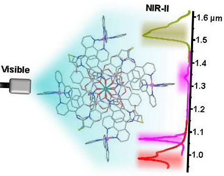
They have overcome this major limitation by designing and synthesising a new family of metallacrowns that have lanthanide sensitizers that can be excited in the visible range of the electromagnetic spectrum. The structure of these metallacrowns is innovative: the sensitizers are attached to the periphery. Thanks to this approach, researchers were able, for the first time, to use a metallacrown to label living cells and image them using near-infrared microscopy. This work opens up major prospects for the use of lanthanide-based molecular complexes for near-infrared imaging in vitro and in vivo.

This major innovation was reported by CNRS Chemistry on its website.

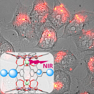
Caption: Near-infrared luminescence image superimposed on the white light image obtained from living HeLa cells in which lanthanide metallacrowns, whose structure appears inlaid, were incubated.

Article references:
Novel lanthanide( III)/gallium( III) metallacrowns with appended visible-absorbing organic sensitizers for
molecular near-infrared imaging of living cells
Timothée Lathion, Julie Bourseguin, Svetlana V. Eliseeva, Matthias Zeller, Stéphane Petoud, Vincent L. Pecoraro
Chemical Science, 2025, 16, 12623. https://doi-org.insb.bib.cnrs.fr/10.1039/D5SC01320H

CBM doctoral student wins prize for best oral presentation at symposium

Petra Cutuk, doctorante au CBM, a reçu le prix de la meilleure présentation orale lors du colloque 2025 du Réseau Molécules Marines, Métabolisme et Cancer.

Petra Cutuk, doctorante de deuxième année, qui effectue son travail de recherche conjointement entre l'équipe "Composés luminescents de lanthanides, spectroscopie et bioimagerie optique" du CBM d'Orléans et l'équipe Inserm "U1069 - Niche, Nutrition, Cancer & métabolisme Oxydatif- N2Cox" de l'Université de Tours, a reçu le prix de la meilleure présentation orale pour sa présentation intitulée "Elucidating the Regulation of SKCa Channels by the cAMP/PKA Pathway in Cancer Cells: Innovations in Near-Infrared Optical Imaging Agents" lors du colloque annuel 2025 du Réseau Molécules Marines, Métabolisme & Cancer à Mansigné le 16 mai 2025.

The prize is awarded by the Fondation ARC pour la recherche sur le cancer to finance participation in a conference.

Petra Cutuk is on the right in the photo.


Multiplex imaging in the NIR-II window with lanthanide-based molecular agents: a dream or a reality?

In this work we have created a new family of lanthanide-based molecular imaging agents that can be used for multiplex imaging in the second near-infrared window (NIR-II, 1000-1700 nm). NIR-II light is particularly interesting for non-invasive, real-time imaging and diagnosis of living organisms, as it is unaffected by the native fluorescence of biological tissues and fluids, and can penetrate through them. In addition, multiplex imaging enables the simultaneous, real-time visualization of several biological markers for even more precise diagnosis of diseases and a deeper understanding of biological processes. This new family of molecular imaging agents is based on an innovative design and combines the exceptional advantages of “metallacrowns” to emit NIR-II light with those of ruthenium complexes, which exhibit strong absorption in the visible range and effectively sensitize lanthanides. Using NIR-II imaging experiments, we have been able to demonstrate that four bands from three different lanthanides can be distinguished unambiguously due to their minimal overlap, while exhibiting sufficient intensity to be detected through a tissue-mimicking phantom.

Enabling Visible Light Sensitization of YbIII, NdIII and ErIII in Dimeric LnIII/GaIII Metallacrowns through Functionalization with RuII Complexes for NIR-II Multiplex Imaging
Codruţa C. Bădescu-Singureanu, Dr. Anton S. Nizovtsev, Prof. Dr. Vincent L. Pecoraro, Prof. Dr. Stéphane Petoud, Dr. Svetlana V. Eliseeva
Angewandte Chemie International Edition 2024
https://doi-org.inc.bib.cnrs.fr/10.1002/anie.202416101

This article was reported by CNRS Chimie on its website.

Professor Vincent L. Pecoraro of the University of Michigan is invited at the CBM from May to July 2024 through a LE STUDIUM Research Professorship

Professor Pecoraro is internationally recognized for his major contributions in bioinorganic chemistry, notably on the role of manganese complexes and clusters in photosynthetic water oxidation. He is also a pioneer in the design of metalloenzyme mimetics and in the study of metallacrown complexes with unique magnetic and/or luminescence properties for developments in medical imaging, a field in which several patents have recently been filed with the team “Luminescent lanthanide compounds, optical spectroscopy and bioimaging” from the CBM.

In 2021, he received an honorary doctorate from the University of Aix-Marseille and a LE STUDIUM Research Professorship (Loire Valley Institute). In addition, Professor Pecoraro is the 2021 recipient of the Franco-American award from the American Chemical Society and the “Société Chimique de France”.

He is one of the co-founder of the startup VIEWaves founded in Orléans in March 2022 that aims at the commercialization of the optical imaging agents emitting in the near-Infrared domain created on the basis of the collaborative work conducted with the team at CBM.

Dual agents for non-invasive imaging of living organisms

Researchers in the "Lanthanide Luminescent Compounds, Spectroscopy and Optical Bioimaging" team have designed a molecular probe that can image living organisms using both near-infrared (NIR) luminescence and photoacoustic (PA) signal detection. These two complementary imaging techniques make it possible to monitor biological events precisely, in real time and non-invasively.

Find out more

A Dual-Mode Near-Infrared Optical and Photoacoustic Imaging Agent Based on a Low Energy Absorbing Ytterbium Complex
Anton Kovalenko, Svetlana V. Eliseeva, Guillaume Collet, Saïda El Abdellaoui, Sharuja Natkunarajah, Stéphanie Lerondel, Laure Guénée, Céline Besnard & Stéphane Petoud
JACS 2024
https://doi.org/10.1021/jacs.4c03406