A CBM team has developed a contrast agent capable of revealing oxidative stress in living tissue

The "Metal Complexes and MRI" team, in collaboration with a Hungarian team, has developed a contrast agent based on an iron compound associated with a fluorinated ligand. This makes it possible to map oxidative stress in living tissue, a marker of numerous pathologies. This molecular detector, still at the pre-clinical stage, could considerably enrich tomorrow's medical imaging toolkit.

This major advance has been published in the journal JACS.
Relaxation-Based In Vivo Discrimination of Oxidized and Reduced States of a Redox-Switchable 19F MRI Probe
Garda Z., Szeremeta F., Tóth C.S., Bunda S., Pifferi C., Clémençon R., Même S., Tircso G., Tóth É.
J. Am. Chem. Soc. 2025, 147, 21, 18017-18024

Find out more on the CNRS Chimie website.


Martina Sanadar, PhD in Environmental and Energy Engineering, is a post-doctoral fellow at CBM

Originally from Italy, Martina Sanadar has a PhD in environmental and energy engineering. She has been a post-doctoral fellow in the "Metal Complexes and MRI" team for the past year. Her research focuses on the development of bioinspired MRI contrast agents sensitive to Cu²⁺. In vivo imaging of extracellular Cu²⁺ is of great interest due to its biological importance in physiological and pathological states. However, the development of effective and selective MRI contrast agents for Cu²⁺, especially compared to the more abundant Zn²⁺ ions, remains a considerable challenge.

Martina Sanadar contributes her expertise in coordination and physical chemistry, solution thermodynamics and various techniques (potentiometry, relaxivity measurements, luminescence spectroscopy, UV-visible spectroscopy and calorimetry).

She joined the CBM for the excellence of its research, its collaborative environment and advanced equipment, as well as the opportunity to work in France. The CBM's interdisciplinary approach and international collaborations align perfectly with her professional goals, making it an ideal destination to further her career.

‘Etonnante Chimie’ (Amazing Chemistry): a CBM chemistry team welcomed secondary school students from Tours

Following the publication of the book “ Etonnante Chimie ” and as part of the project “ Etonnante chimie pour un grand oral percutant ” (“Amazing Chemistry for a Successful Oral Exam”), the “Metal complexes and MRI” team, led by Dr Eva Jakab Toth and Célia Bonnet, has been welcoming, for 4 consecutive years, first-year high school students from the Vaucanson high school in Tours.

Will it be possible to soon detect copper by non invasive imaging?

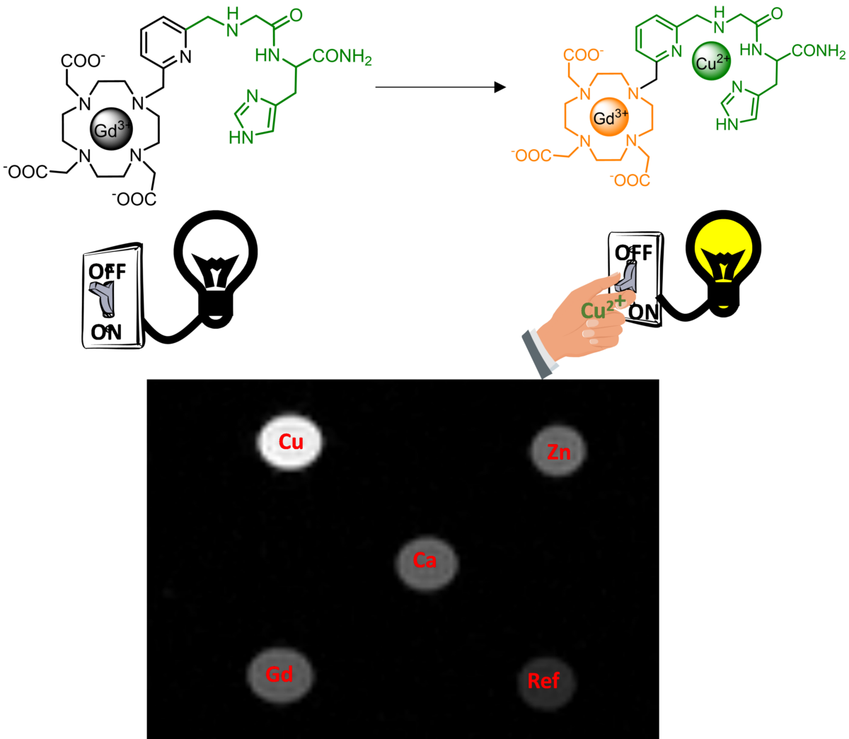
In this work, in collaboration with chemists from the Institut de Chimie de Strasbourg (CNRS/Université de Strasbourg), we have designed and studied a smart MRI probe; which is switched on in the presence of copper. The design of such probes is a real challenge as free Cu(II) in vivo is present in very low quantities, typically lower than Zn(II), another physiological cation. It is therefore of prime importance to conceive probes with a maximal turn on response in the presence of Cu(II), and an excellent selectivity towards Zn(II). The probes are typically composed of an MRI active site, a linker and a Cu(II) binding site. The use of small complexing units for Cu(II) binding makes it very difficult to obtain a good selectivity. Here, we have used a bioinspired approach where the Cu(II) binding site is based on the ATCUN motif, a small peptide that binds Cu(II) in the blood. Thanks to this design, the probe displays an unprecedented turn on response, and importantly an excellent selectivity for Cu(II) vs Zn(II). Phantom MRI images obtained closed to physiological conditions show a bright contrast, illustrating the potential of such probes.

Reference :
A Bioinspired Cu2+-Responsive Magnetic Resonance Imaging Contrast Agent with Unprecedented Turn-On Response and Selectivity
Katharina Zimmeter, Agnès Pallier, Bertrand Vileno, Martina Sanadar, Frédéric Szeremeta, Carlos Platas-Iglesias, Peter Faller, Célia S. Bonnet and Angélique Sour
Inorganic Chemistry - Vol 63 - Issue 49 - 23067−23076

Contrast agents to combine 1H and 19F MRI

Today, magnetic resonance imaging (MRI) is based on the detection of water protons (1H) in tissues. MRI of 19F fluorine offers complementary advantages, but its use is hampered by a lack of suitable imaging agents, soluble in water and easily detectable. In order to improve the sensitivity of detecting 19F MRI signals, a CBM team used Mn2+ ions to form complexes with small fluorinated molecules. Unlike currently used nanoparticles, these small molecular probes have well-defined chemical structures and better biocompatibility and water solubility. Finally, thanks to the paramagnetism of manganese(II), they generate a strong signal in MRI. In addition, these fluorinated contrast agents are also active in proton MRI, allowing proton and fluorine MRI images to be superimposed for precise anatomical mapping.

This advance, published in Angewandte Chemie International Edition, opens new horizons in fluorine MRI. It was reported by CNRS Chimie on its website.

Reference :
Small, Fluorinated Mn2+ Chelate as an Efficient 1H and 19F MRI Probe
Éva Tóth, Zoltán Garda, Frédéric Szeremeta, Océane Quin, Enikő Molnár, Balázs Váradi, Rudy Clémençon, Sandra Même, Chantal Pichon and Gyula Tircsó
Angewandte Chemie International Edition, 2024
DOI: 10.1002/anie.202410998