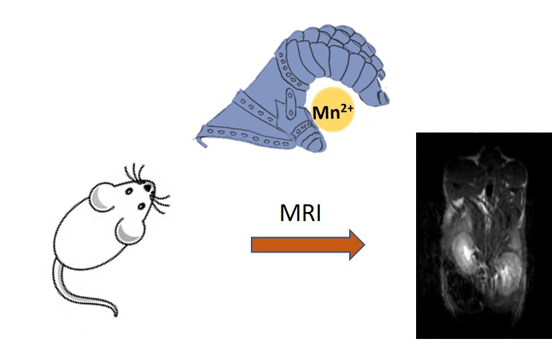
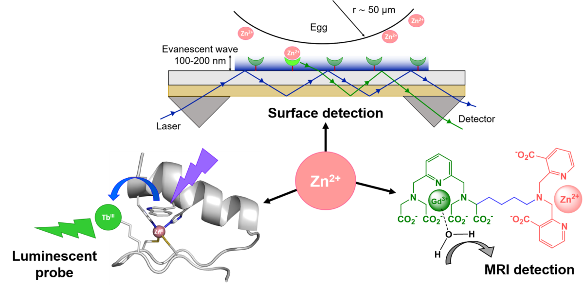
Towards new imaging markers for in vivo detection of pathologies such as Alzheimer’s or diabetes

The accumulation of aggregates of certain non-soluble peptides in tissues is characteristic of several pathologies, such as Alzheimer's and Parkinson's diseases, or diabetes. The detection of these amyloid deposits by in vivo imaging would be very useful for an early diagnostic and a better understanding of the molecular mechanisms of these diseases. Researchers from the Molecular Biophysics Center (CBM) and the Toulouse Coordination Chemistry Laboratory (LCC), in collaboration with Portuguese and Hungarian scientists, have taken an important step forward in the design of imaging agents that specifically recognize these amyloid deposits. This work is on the cover of Chemistry A European Journal.

Read more on the website of CNRS Chemistry Institute (INC)


Detection of amyloid peptides: biomarkers for Alzheimer’s Disease and Diabetes

Metal chelates targeted to amyloid peptides are widely explored as diagnostic tools or therapeutic agents for amyloidogenic diseases. For example, gadolinium complexes can be used as MRI probes, while radiocomplexes (64Cu, 99mTc, etc) can be exploited for nuclear imaging. Other metal complexes capable of preventing aggregate formation are proposed to derive therapeutic strategies.

All these molecules are amphiphilic, composed of a hydrophilic part (containing the metal) and a hydrophobic one (capable of targeting the amyloid aggregates). This particular structure enables the formation of micelles in solution.

The groups of CBM and LCC (Toulouse) have found that this micellisation property has drastic and unexpected consequences on their ability to recognize amyloid peptides and a high impact on their in vivo biodistribution.

The researchers have developed a novel probe with nanomolar affinity towards Aβ and amylin, biomarkers of Alzheimer’s Disease and Ddiabetes, respectively.

This exceptional affinity for a metal complex is only obtained if the complex is present as “single molecule”. Once in the micellar form, this affinity drops by 1000-fold.

These results have a direct consequence for the design of novel imaging and therapeutic probes for amyloidogenic pathologies.

See the article

Manganese: A double agent for imaging ?

Gadolinium (Gd3+) complexes have been used as MRI contrast agents for 35 years, but recently the safety of some was questioned. The replacement of Gd3+ by manganese (Mn2+), a biogenic metal, would enable safer complexes.

Nevertheless, the Mn2+ has to be chelated by complexes exhibiting high thermodynamic stability and kinetic inertness (to guarantee that the Mn is not released in vivo) and with a water molecule directly coordinated to the metal, essential for a good MRI efficiency. Combining these two properties is a chemistry challenge.

The “Metal complexes and MRI” team of CBM and their collaborators from IPHC (Strasbourg) have synthesized and studied a bispidine ligand, a molecule which cavity is well adapted for Mn2+ complexation. This Mn2+ complex has an excellent kinetic inertness and its MRI efficiency was validated by preclinical studies.

52Mn is an emergent radionuclide for positron emission tomography (PET). Mn2+ is the only metal enabling both MRI and PET imaging. The use of 52Mn is nevertheless limited by its low availability and lack of appropriate ligand.

For the first time in France, 52Mn was produced at the Orléans’ cyclotron, and 52Mn-bispidine was successfully obtained.

Overall, bispidine is a very promising ligand for the Mn2+ complexation, for MRI and PET. Due to its outstanding kinetic inertness, in vivo use of Mn2+ without toxicity risk can be anticipated.


See the news on the website of the CNRS Institute for Chemistry.


Eva Toth, Daouda Ndiaye, Maryame Sy, Agnès Pallier, Sandra Même, Isidro de Silva, Sara Lacerda, Aline M. Nonat, Loïc J. Charbonnière Unprecedented kinetic inertness for a Mn2+‐bispidine chelate: a novel structural entry for Mn2+‐based imaging agents - Angewandte Chemie, 2020, https://doi.org/10.1002/anie.202003685

DECLICS

On January 17, 2020, rich exchanges took place between the thematic group "Metal Complexes and MRI" and students of the first and final European class of the Lycée Pothier in Orleans as part of DECLICS "Dialogues Entre Chercheurs et Lycéens pour les Intéresser à la Construction des Savoirs" .

13th Workshop of the “Vectorization, Imaging and Radiotherapy” network of Cancéropôle Grand Ouest – Le Bono, 2019, September 25-28th

The 13th international workshop edition will focus on the latest advances in imaging techniques: PET, SPECT, MRI, US, Infra-red Bioluminescence, Fluorescence, Photoacoustic, CyTof Imaging, Microscopy, Spectroscopy and Raman Imaging.

It aims to address innovative developments in chemistry (synthesis of tracers, chelates, contrast agents, fluorescent probes, etc.), biology (identification of new tumour targets, etc.) and physics (instrumentation, methodological aspects of image analysis, radiomics ...)

The workshop will confront the points of view and problems of the various disciplines, Chemistry, Biology, Physics, Medicine, working for the development of imaging in Oncology.

Overview lectures will be given by experts in the different scientific fields targeted by the workshop. A number of selected oral presentations given by young researchers will complete the different scientific sessions.

This workshop aims at sharing scientific results and at facilitating exchanges between experienced and young researchers in a fast advancing field. The place of the congress was chosen in order to stimulate intense discussions between young and senior scientists.

See workshop site