Séminaire de Manon Isaac, post-doctorante dans le groupe thématique "Complexes métalliques et IRM".
Abstract:
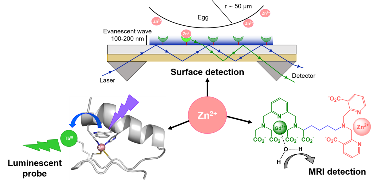
Zinc cations play a key role as a cofactor in gene transcription and metalloenzyme functions, but also in signaling pathways in the immune system, or during fertilization. To understand its underlying function, on top of the quantification of the total amount of zinc, the detection of the labile pool and its evolution is crucial. Whatever the technique is, probes are required to visualize labile zinc. The probes have to be optimized for a given technique but also for the biological context (e.g. extracellular vs intracellular).
Manon Isaac will present several tools to detect labile zinc developed during her PhD and postdocs: design and characterization of peptidic luminescent probes, the first steps towards a device to detect zinc released by the egg after in vitro fertilization, and MRI zinc probes.