Typologie d'actualités: Team Chemistry, Imaging and Exobiology
Searching for the earliest traces of life in the Solar System
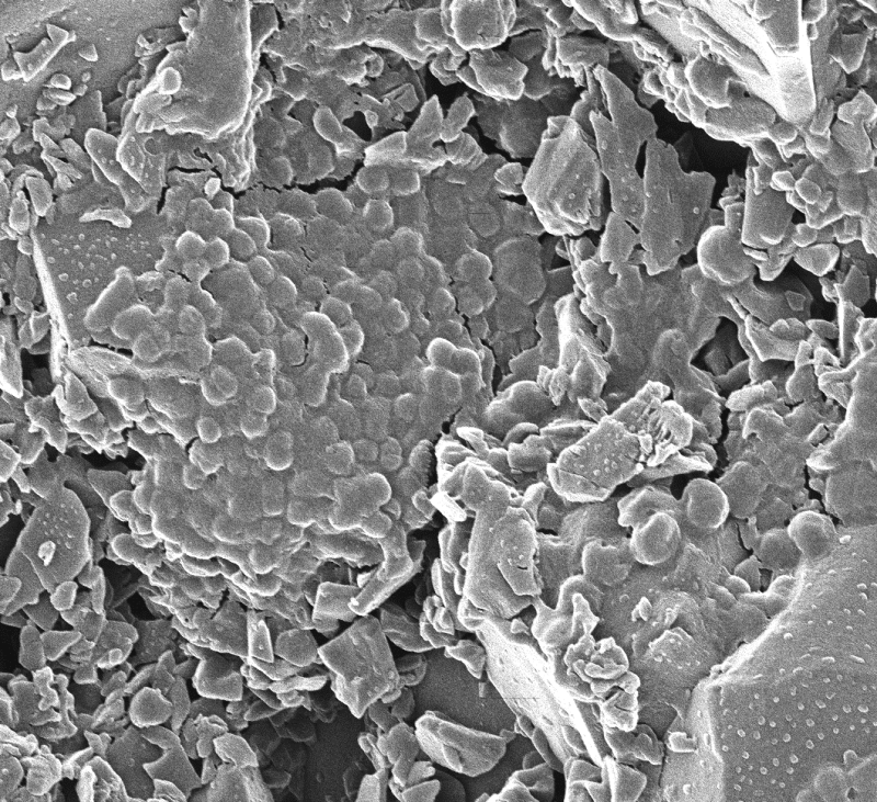
Life appeared on Earth very early on, at a time when our planet was just a hot world bombarded by ultraviolet radiation. These conditions, which were undoubtedly common to other rocky planets such as Mars, may have favored the emergence of simple life forms: microbes that fed on and drew their energy solely from the oxidation of mineral matter. With this in mind, a team from the Center for Molecular Biophysics in Orleans (CNRS), in collaboration with Newcastle University, revisited an iconic and well-preserved site in northwestern Australia, Kitty's Gap Chert, formed in 3.45-billion-year-old coastal volcanic sediments.
Read more on the CNRS Chemistry website.
Article reference:
Insights from early life in the 3.45-Ga Kitty's Gap Chert for the search for elusive life in the Universe
Frances Westall, Graham Purvis, Naoko Sano, Jake Sheriff, Laura Clodoré, Frédéric Foucher & Tetyana Milojevic
Nature Astronomy 2025
https://doi.org/10.1038/s41550-025-02661-0
Stronger RNA building blocks for tomorrow’s vaccines
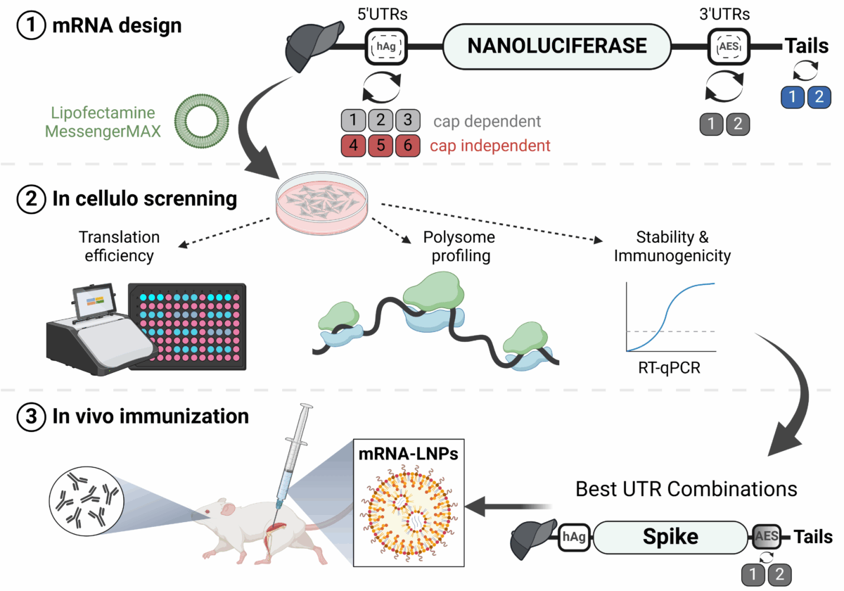
Messenger RNA (mRNA) has emerged as an attractive new technology of drugs. The efficacy of mRNA technology depends on both the efficiency of mRNA delivery and translation. Untranslated regions (UTRs) and the poly(A) tail play a crucial role in regulating mRNA intracellular kinetics. Intending to improve the therapeutic potential of synthetic mRNA, CBM researchers evaluated various UTRs and tail designs, using Pfizer-BioNTech COVID-19 vaccine sequences as a reference. First, they screened six 5’ UTRs (capdependent/independent), evaluated nine 5’ UTR-3’ UTR combinations, and a novel heterologous A/G tail in cell models, and in vivo using luciferase as a reporter gene.
Then, to decipher the translation mechanism of selected UTRs, they correlated mRNA expression with ribosome load, mRNA half-life, mRNA immunogenicity, and UTR structures. Results showed that the heterologous tail they introduced is as potent as the Pfizer-BioNTech tail and confirmed the high potency of the human α-globin 5’ UTR.
They also revealed the potential of the VP6 and SOD 3’ UTRs. Researchers validated their results using mRNA encoding the SARS-CoV-2 spike protein formulated as lipid nanoparticles (LNPs) for mouse immunization. Overall, the selected 3’ UTRs and heterologous A/G tail have great potential as new elements for therapeutic mRNA design.
These results open up new prospects for mRNA therapies. While improvements are still needed to achieve higher expression than existing strategies, this strategy contributes to improving mRNA therapies.
These results are linked to a patent
These major advances were reported by CNRS Chemistry in its scientific news.
Reference:
Evaluation of synthetic mRNA with selected UTR sequences and alternative Poly(A) tail, in vitro and in vivo.
Medjmedj A, Genon H, Hezili D, Ngalle Loth A, Clemençon R , Guimpied C, Mollet L, Bigot A, Wien F, Hamacek J, Chapat C & Perche F
Molecular Therapy Nucleic Acids 2025. DOI: 10.1016/j.omtn.2025.102648
Luminous crowns to see into the heart of life
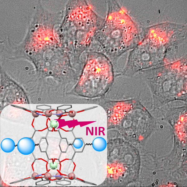
The members of the ‘Luminescent Lanthanide Compounds, Optical Spectroscopy and Bioimaging’ team have been working for many years on the design and synthesis of supramolecular complexes based on lanthanides emitting in the near infrared, which function as optical imaging agents for biological experimentation and medical diagnosis. They have designed lanthanide-containing metallacrown complexes that have proven to be very promising candidates due to their very high brightness. However, until now, these metallacrowns had a significant limitation due to their excitation wavelengths, which were limited to the ultraviolet part of the electromagnetic spectrum. These wavelengths pose a problem for biological imaging because they can severely disrupt or damage the biological system being observed.
They have overcome this major limitation by designing and synthesising a new family of metallacrowns that have lanthanide sensitizers that can be excited in the visible range of the electromagnetic spectrum. The structure of these metallacrowns is innovative: the sensitizers are attached to the periphery. Thanks to this approach, researchers were able, for the first time, to use a metallacrown to label living cells and image them using near-infrared microscopy. This work opens up major prospects for the use of lanthanide-based molecular complexes for near-infrared imaging in vitro and in vivo.
This major innovation was reported by CNRS Chemistry on its website.
Caption: Near-infrared luminescence image superimposed on the white light image obtained from living HeLa cells in which lanthanide metallacrowns, whose structure appears inlaid, were incubated.
Article references:
Novel lanthanide( III)/gallium( III) metallacrowns with appended visible-absorbing organic sensitizers for
molecular near-infrared imaging of living cells
Timothée Lathion, Julie Bourseguin, Svetlana V. Eliseeva, Matthias Zeller, Stéphane Petoud, Vincent L. Pecoraro
Chemical Science, 2025, 16, 12623. https://doi-org.insb.bib.cnrs.fr/10.1039/D5SC01320H
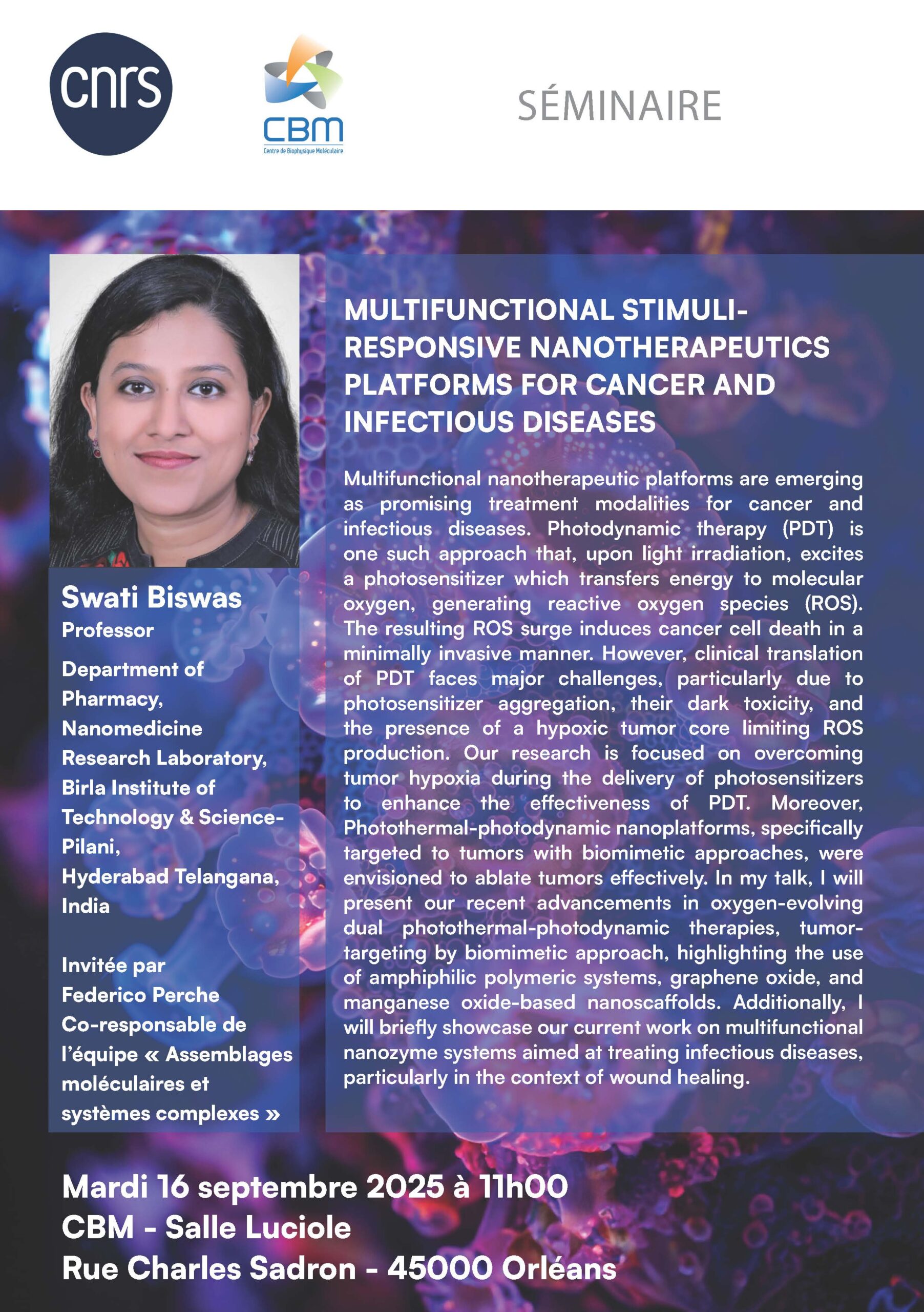
2025, September 16: Swati Biswas seminar

The CBM is recruiting!

Join the “Metal Complexes and MRI” team, co-directed by Drs Eva Jakab Toth and Célia Bonnet.
Consult the job profile and apply.
Starting date: October 1, 2025
Application deadline: Saturday August 9, 2025 23:59:00 Paris time
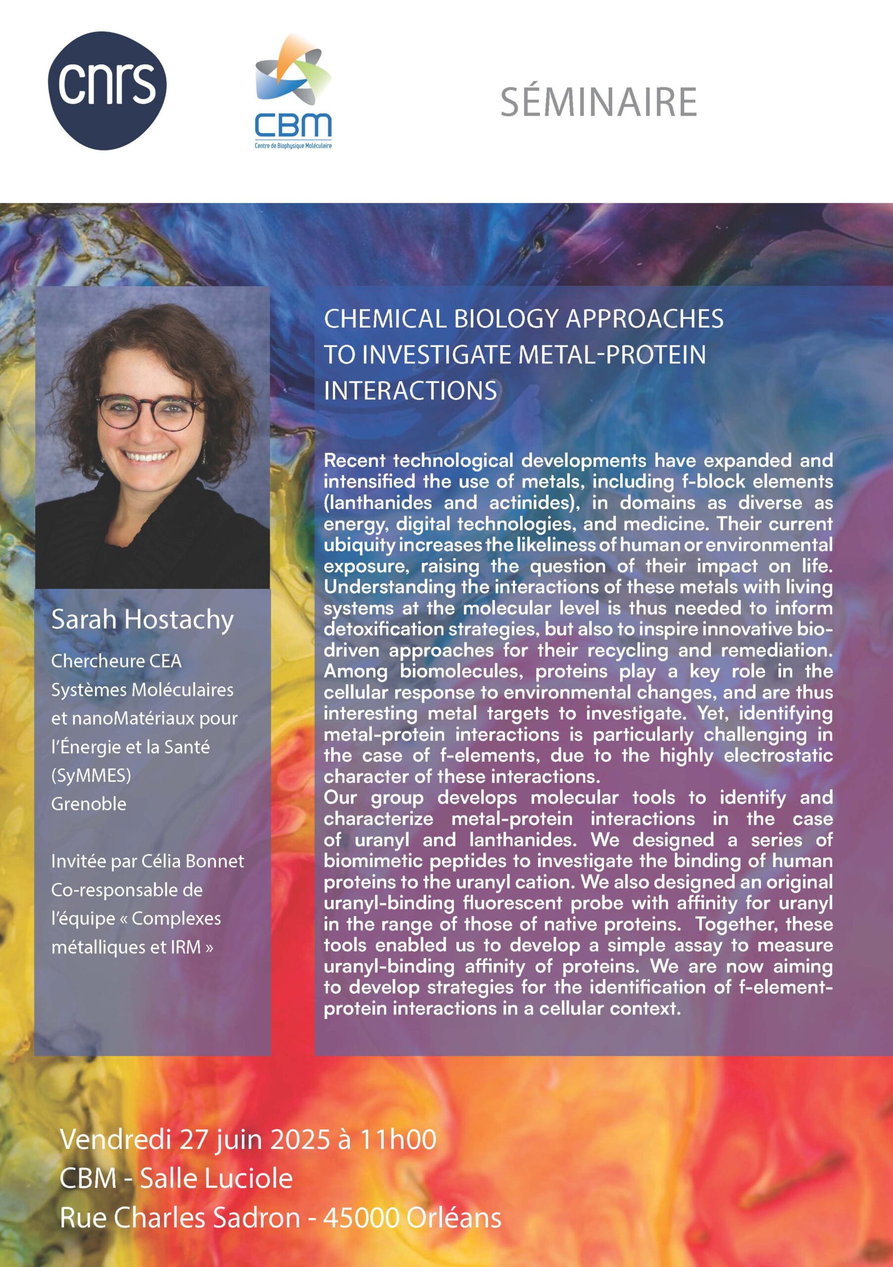
2025, June 25: Sarah HOSTACHY seminar


