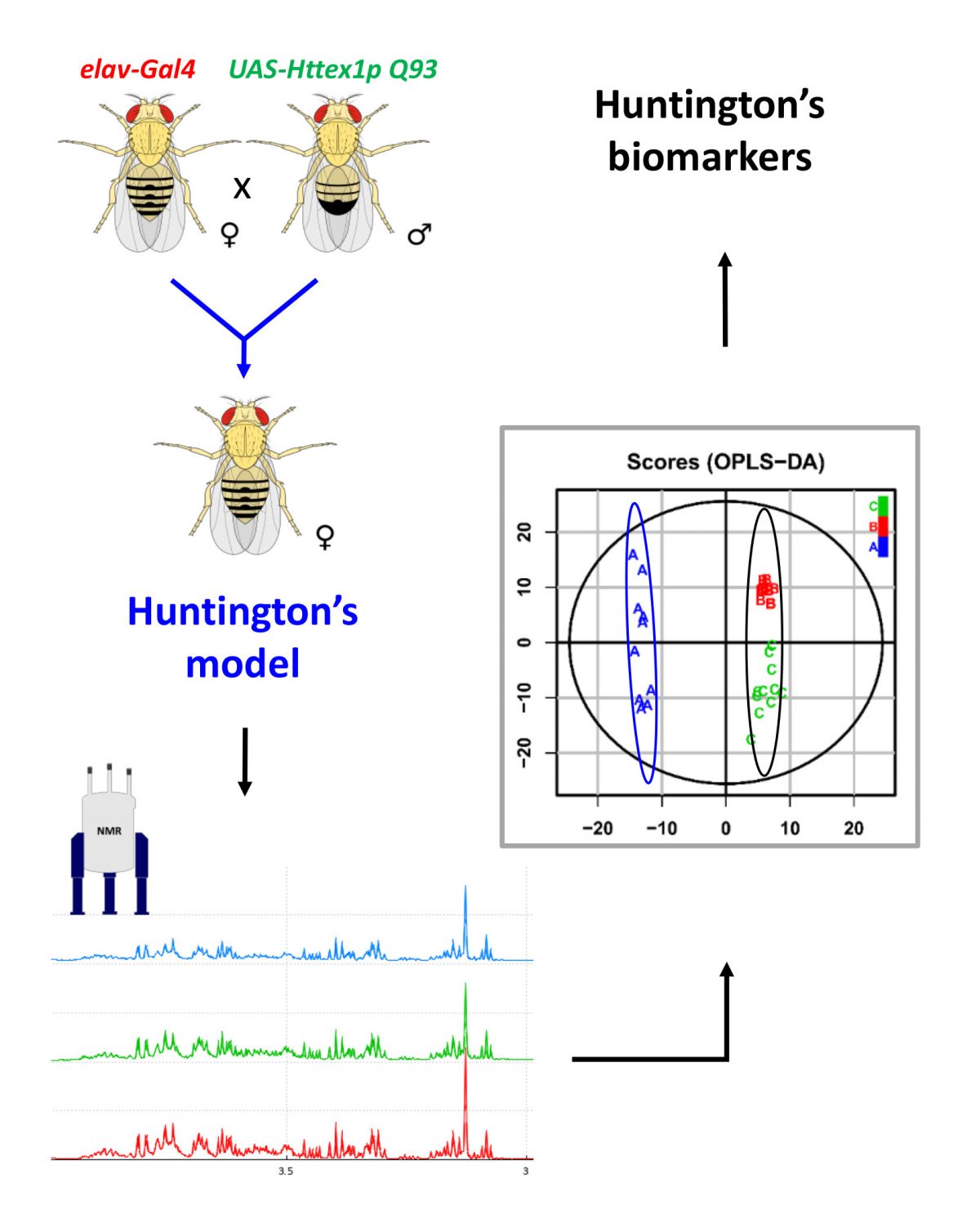
Targeting the LINGO-1 receptor and its hetero-complexes: new therapeutic perspectives for the treatment of demyelinating and neurodegenerative diseases

LINGO-1 is a transmembrane protein mainly expressed in neurons and oligodendrocytes where it negatively regulates regeneration, neuronal survival, oligodendrocyte differentiation and myelination. Its expression was shown to be dysregulated in several brain disorders, in particular, Parkinson's disease and multiple sclerosis, which makes LINGO-1 a particular interesting therapeutic target. Three homologs LINGO-2, LINGO-3 and LINGO-4 have been described in human but until now their functions remain largely unknown. In this article published in the FASEB Journal, the researchers evaluated the expression, the cerebral distribution and the colocalization of these different homologs in several brain areas. They also showed the formation of hetero-complexes of LINGO-1 with its homologs in the brain, revealing a new level of complexity in the regulation of the activity of these receptors. The demonstration of this LINGO-1 signaling opens the way to new strategies to modulate its activity for the treatment of brain disorders.

Guillemain, A., Y. Laouarem, L. Cobret, D. Stefok, W. Chen, S. Boch, A. Zahaf, L. Blot, F. Reverchon, T. Normand, M. Decoville, C. Grillon, E. Traiffort and S. Morisset (2020). "LINGO family receptors are differentially expressed in mouse brain and forms native multimeric complexes " FASEB J (2020) doi : 10.1096/fj.202000826R

Pure and stable white light

Current white light sources, such as LEDs, tend to oscillate between different color variations, which distorts the perception of the human eye. This limitation can be very disabling, even dangerous, during surgery or in the work of graphic designers and artists.

Researchers from the Center for Molecular Biophysics and the University of Michigan (United States) have developed a new system, based on dysprosium atoms and metallacrowns, which gives exactly white light that does not vary with conditions wear or temperature.

This work has been published in the Journal of the American Chemical Society.

See the information on the CNRS Institute of Chemistry website.

Does your ten-year-old code still run?

Computers have become indispensible tools in scientific research, and software is increasingly becoming a medium for expressing scientific models and methods. But contrary to journal articles, which are archived and remain accesssible for many decades, software is fragile and can become unusable within a few years.

CBM researcher Konrad Hinsen and computer scientists Nicolas Rougier from INRIA Bordeaux Sud-Ouest have launched a challenge to computational scientists: Can you still run code that you published at least ten years ago?

In response, the journal ReScience C, which they founded in 2015, received 28 submissions of detailed reproducibility reports, which have been summarized by a journalist for Nature.

Creation of a predictive model of metabolic changes characteristic of neurodegenerative diseases

Drosophila flies are versatile animal models for the study of genetic mutations in neuronal pathologies. Their small size allows performing in vivo experiments to obtain high resolution nuclear magnetic resonance spectra. Dr. Martine Decoville of CBM, researchers from CEMHTIand ESPCI Paris used spatially-resolved ¹H high-resolution MAS NMR to investigate in vivo metabolite contents in different segments of the fly body. A comparative study of metabolic changes was performed for three neurodegenerative disorders: two cell-specific neuronal and glial models of Huntington disease and a model of glutamate excitotoxicity. These 3 pathologies are characterized by specific and sometimes anatomically localized variations in metabolite concentrations. In two cases, the modifications of ¹H MAS NMR spectra localized in fly heads were significant enough to allow the creation of a predictive model.

Spatially-resolved metabolic profiling of living Drosophila in neurodegenerative conditions using 1H magic angle spinning NMR - Scientific Reports (2020) https://doi.org/10.1038/s41598-020-66218-z

Disturbed expression of autophagy genes in blood of Parkinson’s disease patients

Parkinson's disease is a neurodegenerative pathology characterized by the presence of protein aggregates in the neurons of patients. This neurotoxic accumulation of misfolded proteins could be due to insufficient elimination by autophagic cellular mechanisms. Alain Legrand's group has just published a study highlighting an alteration in the expression of genes coding for autophagy proteins in the blood of patients with Parkinson's disease. This pilot study, carried out in collaboration with the Neurology Department of the Centre Hospitalier Régional d'Orléans (CHRO) and the Fundamental Informatics Laboratory of the University of Orléans (LIFO), shows that defects in autophagic systems that affect the brain of patients can also be detected in peripheral blood cells. These deregulated genes could thus constitute diagnostic markers of Parkinson's disease that can be measured non-invasively after a simple blood test.

El Haddad S. et al, Disturbed expression of autophagy genes in blood of Parkinson's disease patients.  Gene vol. 738 (2020): 144454

Unveiling billion-year old life forms with X-ray vision

An international team of scientists from Brazil, France and Switzerland with financial support from the Serrapilheira Institute and Fapesp, has obtained the most detailed 3D views ever achieved of very ancient traces of life on Earth. The studied microfossils, from the Gunflint Formation, in Canada, are approximately 1.9 billion years old, and are the preserved remains of microorganisms similar to bacteria existing today, but from a period when only microscopic life existed on Earth. Using an advanced imaging method based on synchrotron light, unprecedented details of the shape, composition and preservation of these microfossils was attained. Moreover, in one locality, fossils previously termed “hematite-coated” are revealed to be composed of organic material – invisible in optical microscopy – coated with crystals of the iron oxide maghemite, instead of hematite. This finding challenges our understanding of past life and opens exciting perspectives for the study of even older fossils or future samples returned from Mars.

Maldanis, L., Hickman-Lewis, K., Verezhak, M. et al. Nanoscale 3D quantitative imaging of 1.88 Ga Gunflint microfossils reveals novel insights into taphonomic and biogenic characters. Scientific Reports 10, 8163 (2020). https://doi.org/10.1038/s41598-020-65176-w

Read the article

3D observation of microfossils

Organometallic networks for the near-infrared emission of lanthanides

Researchers from the group "Luminescent lanthanide compounds, spectroscopy and optical bioimaging" and the University of Pittsburgh (USA) have designed a rigid three-dimensional chemical system of organometallic network type composed of lanthanides and organic molecules. A major originality of this work is based on the use of the cavity of this network to generate in situ the molecular system sensitizing the lanthanide cations. This new approach has the advantage, among other things, of allowing the excitation length to be controlled. This network is well suited to the real conditions of optical biomedical imaging on living cells.

See the news on the CNRS website

These researches have been published on Journal of American Chemical Society

Patrick F. Muldoon, Guillaume Collet, Svetlana V Eliseeva, Tian-Yi Luo, Stephane Petoud, and Nathaniel L Rosi. Ship-in-a-bottle preparation of long wavelength molecular antennae in lanthanide metal-organic frameworks for biological imaging. J. Am. Chem. Soc. (2020) 142, 8776-8781 - doi : 10.1021/jacs.0c01426