Manganese: A double agent for imaging ?

Gadolinium (Gd3+) complexes have been used as MRI contrast agents for 35 years, but recently the safety of some was questioned. The replacement of Gd3+ by manganese (Mn2+), a biogenic metal, would enable safer complexes.

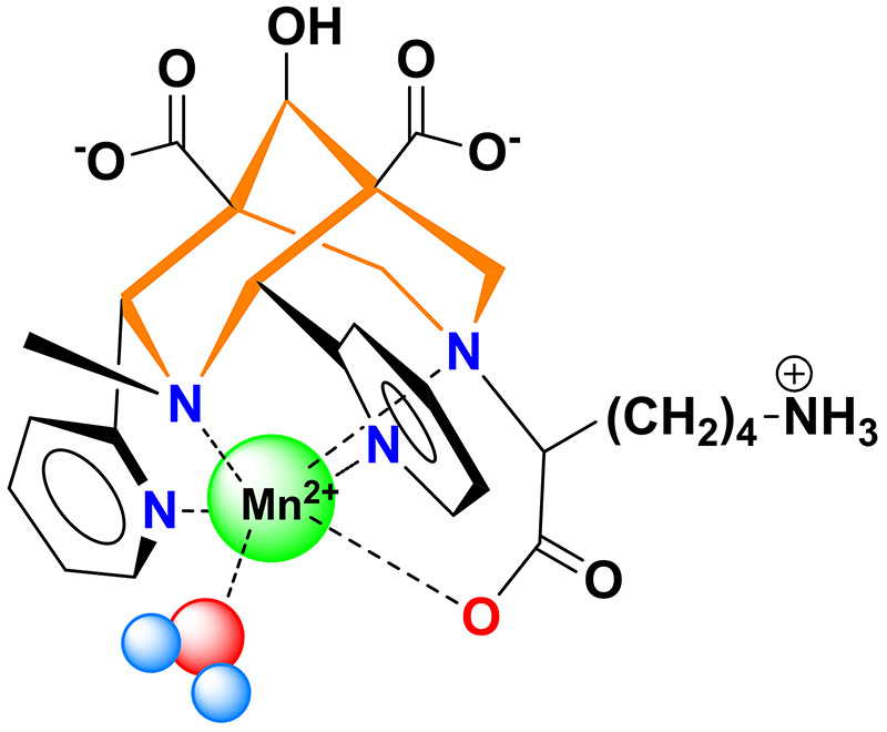
Nevertheless, the Mn2+ has to be chelated by complexes exhibiting high thermodynamic stability and kinetic inertness (to guarantee that the Mn is not released in vivo) and with a water molecule directly coordinated to the metal, essential for a good MRI efficiency. Combining these two properties is a chemistry challenge.

The “Metal complexes and MRI” team of CBM and their collaborators from IPHC (Strasbourg) have synthesized and studied a bispidine ligand, a molecule which cavity is well adapted for Mn2+ complexation. This Mn2+ complex has an excellent kinetic inertness and its MRI efficiency was validated by preclinical studies.

52Mn is an emergent radionuclide for positron emission tomography (PET). Mn2+ is the only metal enabling both MRI and PET imaging. The use of 52Mn is nevertheless limited by its low availability and lack of appropriate ligand.

For the first time in France, 52Mn was produced at the Orléans’ cyclotron, and 52Mn-bispidine was successfully obtained.

Overall, bispidine is a very promising ligand for the Mn2+ complexation, for MRI and PET. Due to its outstanding kinetic inertness, in vivo use of Mn2+ without toxicity risk can be anticipated.


See the news on the website of the CNRS Institute for Chemistry.


Eva Toth, Daouda Ndiaye, Maryame Sy, Agnès Pallier, Sandra Même, Isidro de Silva, Sara Lacerda, Aline M. Nonat, Loïc J. Charbonnière Unprecedented kinetic inertness for a Mn2+‐bispidine chelate: a novel structural entry for Mn2+‐based imaging agents - Angewandte Chemie, 2020, https://doi.org/10.1002/anie.202003685

Metallomics in geological time: trace element biosignatures evidence the influence of ocean chemistry on Earth’s earliest ecosystems

We used a combination of techniques: microbeam particle-induced X-ray emission spectroscopy (PIXE), carbon isotope geochemistry and electron microscopy. This has allowed us to discover trace element signatures of life in 3.33 billion-year-old rocks from South Africa. These signatures support a long-standing hypothesis that biological dependency on trace elements results from the enrichment of these elements in the metal-rich, hydrothermally influenced habitats of early life.

We approached this challenge through the biological concept of the metallome, which refers to the entirety of the inorganic species (metal and metalloid) within a cell. Although the genome and proteome do not survive fossilisation over billions of years, it is probable that metal concentrations within carbonaceous materials could do so, and indeed we found this to be the case in numerous carbon-rich microstructures from the Josefsdal Chert.

We found that a range of elements crucial to anaerobic microbes, including Fe, V, Ni, As and Co, were enriched within carbonaceous material characterised by negative carbon isotope signatures indicating biological origins.

Palaeo-metallome compositions could be used to deduce the metabolic networks of Earth’s earliest ecosystems and, potentially, as a biosignature for the evaluation of organic materials found on Mars.

The article “Metallomics in deep time and the influence of ocean chemistry on the metabolic landscapes of Earth’s earliest ecosystems” released March 18th in Scientific Reports.

Contact: keyron.hickman-lewis@cnrs.fr; frances.westall@cnrs.fr


Numerical simulation to better select drugs before clinical trials

Chemists from the Institute of Organic and Analytical Chemistry (ICOA, CNRS / University of Orléans) and the Center for Molecular Biophysics (CBM, CNRS) propose a new in silico model, which describes the duration of interactions between a molecule and its biological target. Published in the Journal of Chemical Information and Modeling, this work has successfully predicted effects on a protein linked to certain cancers and helps to reduce doses and thus toxicity.

See the communication from the CNRS Institute for Chemistry

Antimicrobial peptides: an atypical double-domain avian defensin, specifically found in eggs, reveals multiple roles in protection of the embryo

Read CNRS Press release

Gga-AvBD11, the avian β-defensin 11 of the common chicken Gallus gallus (Gga-AvBD11), is egg-specific, and represents the sole double-sized defensin (9.3 kDa) among the 14 AvBDs reported in the chicken species. The appearance of such a double-domain protein during evolution could be driven either by its increased biological potency compared to a single domain molecule, and/or by the necessity to acquire new functions carried only by the full-length protein. To assess the contribution of the two domains, we chemically synthesized them. We determined the 3D NMR structure each domain, and the structure of the compact full-length Gga-AvBD11, composed of two packed β-defensin domains. There is no reference for such a double-β-defensin in structural databanks. Thus, AvBD11 is the archetype of a new structural family, which we named avian-double-β-defensins (Av-DBD).
Its high sequence conservation among birds suggests its essential roles in the avian egg. In collaboration with several teams (Nouzilly and Tours, France), we showed that Gga-AvBD11 displays antimicrobial activities against Gram+ and Gram- bacterial pathogens, the avian protozoan Eimeria tenella and avian influenza virus (H1N1). It also shows cytotoxic and anti-invasive activities, suggesting that it may be involved in the (re-)modeling of embryonic tissues. Our results point to a critical importance of the cationic N-ter domain in mediating antibacterial, antiparasitic and anti-invasive activities, with the C-ter domain potentiating the two latter activities. Strikingly, antiviral activity in infected chicken cells requires the full-length protein.

The benefit for the avian species of possessing a double-sized defensin is a fascinating question. In order to better understand the structure-activity and phylogenetic relationships of AvBD11s family, we are currently studying other AvBD11 proteins (SAPhyR-11 project grant from the Région Centre Val de Loire).

This work was funded by the MUSE (Grant no. 2014-00094512) and SAPhyR-11 (Grant no. 2017-119983) project grants from the Région Centre-Val de Loire.

Structure, function, and evolution of Gga-AvBD11, the archetype of the structural avian-double-β-defensin family
Nicolas Guyot, Hervé Meudal, Sascha Trapp, Sophie Iochmann, Anne Silvestre, Guillaume Jousset, Valérie Labas, Pascale Reverdiau, Karine Loth, Virginie Hervé, Vincent Aucagne, Agnès F. Delmas, Sophie Rehault-Godbert, and Céline Landon

Antimicrobial peptides: How peptide chemistry and NMR shed light on the antimicrobial activity of big defensins

Big defensins, ancestors of β-defensins, are composed of a β-defensin-like C-terminal domain and a hydrophobic ancestral N-terminal domain.

This unique structure is found in a limited number of phylogenetically distant species, mostly living in marine environments. Using solid phase peptide chemistry and native chemical ligation, we produced the oyster Crassostrea gigas BigDef1 (Cg-BigDef1) and its separate domains and characterized their 3D structure by NMR. Cg-BigDef1 showed salt-stable and broad-range bactericidal activity, including against multidrug-resistant clinical isolates of S. aureus. We found that the ancestral N-terminal domain confers salt-stable antimicrobial activity to the β-defensin-like domain, which is otherwise inactive. Moreover, upon contact with bacteria, the N-terminal domain drives Cg-BigDef1 assembly into nanonets that entrap and kill bacteria. We speculate that the hydrophobic N-terminal domain of big defensins has been retained in marine phyla to confer salt-stable interactions with bacterial membranes in environments where electrostatic interactions are impaired.

Those remarkable properties open the way to future drug developments when physiological salt concentrations inhibit the antimicrobial activity of vertebrate β-defensins (ANR MOSAR-Def 2019-2023).

Many thanks to D. Destoumieux-Garzón for collaboration, to “Vaincre La Mucovidose“ and CNRS PEPS X-life” for funding.

Read the article in mBio

Control of bacterial virulence by transcription factors NusG and Rho

The virulence genes of pathogenic enterobacteria are concentrated in genomic islands acquired by horizontal transfer during evolution. The expression of these genes outside the infection phase is detrimental to the bacterium and is therefore highly regulated. A major regulatory mechanism relies on the histone-like protein H-NS, which binds to AT-rich sites characteristic of horizontally acquired DNA and forms oligomeric structures that inhibit transcription over extended regions. These regions, however, remain exposed to invasive transcription from neighboring regions or to H-NS repression defects. Our data support a model in which the transcription elongation factor NusG “secures” the inhibition of virulence genes by stimulating the activity of the transcription termination factor Rho in regions silenced by H-NS. Remarkably, NusG changes the specificity of the Rho factor, which alone preferentially targets C-rich regions. The perturbation of this NusG/Rho-dependent mechanism in Salmonella has profound physiological consequences, probably because unstopped transcription in H-NS -targeted regions feeds a feed-forward activation cascade leading to the uncontrolled expression of pathogenicity islands and co-regulated loci.

Link to the article

Everything you’ve always wanted to know about protein/protein interactions and kinase activity assays is in a paper published in JOVE.

"Cell signalling" group led by the Dr H. Bénédetti has published a video paper in JOVE, Journal of Visualized Experiments, a peer-reviewed scientific journal that publishes experimental methods in video format.

This paper belongs to the Method section focusing on basic technics in Biochemistry.

Different experiments are detailled:

- Transient cell transfections
- Protein extraction
- Study of protein/protein interaction by co-immunoprecipitation experiments
- Study of kinase activity by g32P-[ATP] labeling or by using phospho-specific antibodies

All these different technics are illustrated with data obtained by the group on LIMK2-1, a new protein the team has just pointed out and characterized. LIMK2-1 protein does exist, and it is very atypical in the way it regulates actin cytoskeleton remodeling.