Although every cell in our body contains the same genetic information, cells differ in the way they use it, a process known as “gene expression”. The regulation of gene expression is orchestrated by proteins called transcription factors, which bind to specific sequences within DNA. Transcription factors are traditionally thought to operate mainly as single molecules or dimers.
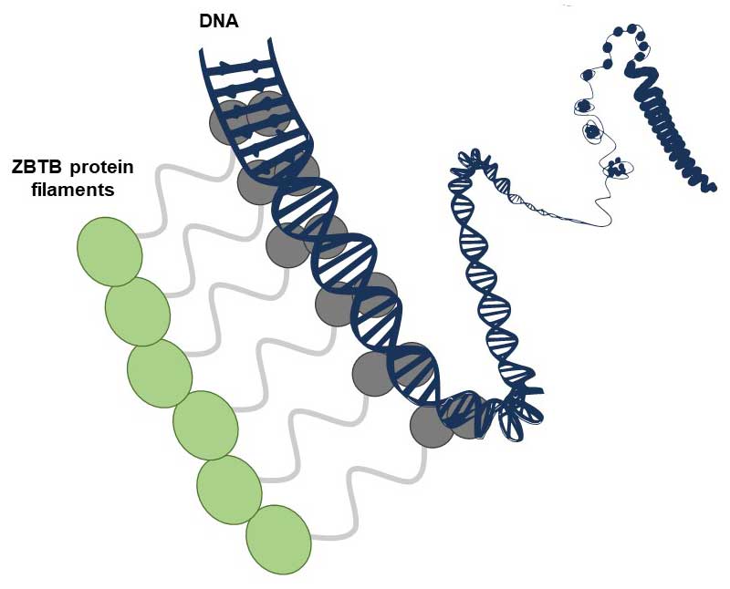
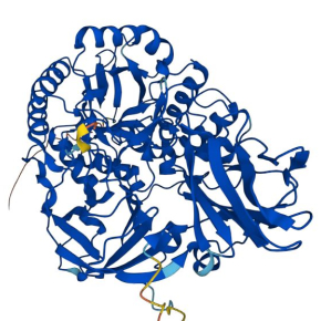
The article by Mance et al. reveals that several transcription factors of the family known as ZBTB, present in humans and other animals, have the capacity to form non-covalent filamentous structures composed of numerous identical copies of proteins arranged in a chain. At the molecular level, such structures could offer significant advantages for binding to DNA, which is itself an elongated molecule containing numerous repeated sequences. A few examples of filament-forming transcription factors had already been reported, but this study extends the concept to a large family of this protein with important functions. The study - which combines structural, biophysical and functional analysis carried out in vitro and in cells - was carried out by the "Post-translational modifications and DNA repair" team at the CBM and their collaborators in Orléans, Rennes and Marseille, including the "Functional mass spectrometry of molecular assemblies" team also at the CBM.
The findings from this research, together with a complementary study by groups of Benjamin Ebert and Eric Fischer from Dana-Farber Cancer Institute at Harvard (published back-to-back in the same issue of Molecular Cell), challenge the traditional view of transcription factor functionality.
In cells, ZBTB proteins are regulated through a process called SUMOylation, where a small tag called SUMO is added to them, changing how they function. Studies on ZBTB proteins undertaken in Orléans, during which the filamentous structures were discovered, are part of the "SUMOwriteNread" project funded by the European Union (ERC grant no 101078837). Researchers are currently investigating the interplay between the ability to form filaments and SUMO tagging to understand the complex reality of gene expression regulation.
This research was reported by CNRS Chimie on its website.
Dynamic BTB-domain filaments promote clustering of ZBTB proteins.
Lucija Mance, Nicolas Bigot, Edison Zhamungui Sánchez, Franck Coste, Natalia Martín-González, Siham Zentout, Marin Biliškov, Zofia Pukało, Aanchal Mishra, Catherine Chapuis, Ana-Andreea Arteni, Axelle Lateur, Stéphane Goffinont, Virginie Gaudon, Ibtissam Talhaoui, Ignacio Casuso, Martine Beaufour, Norbert Garnier, Franck Artzner, Martine Cadene, Sébastien Huet, Bertrand Castaing & Marcin Józef Suskiewicz
Molecular Cell 2024
https://doi.org/10.1016/j.molcel.2024.05.029