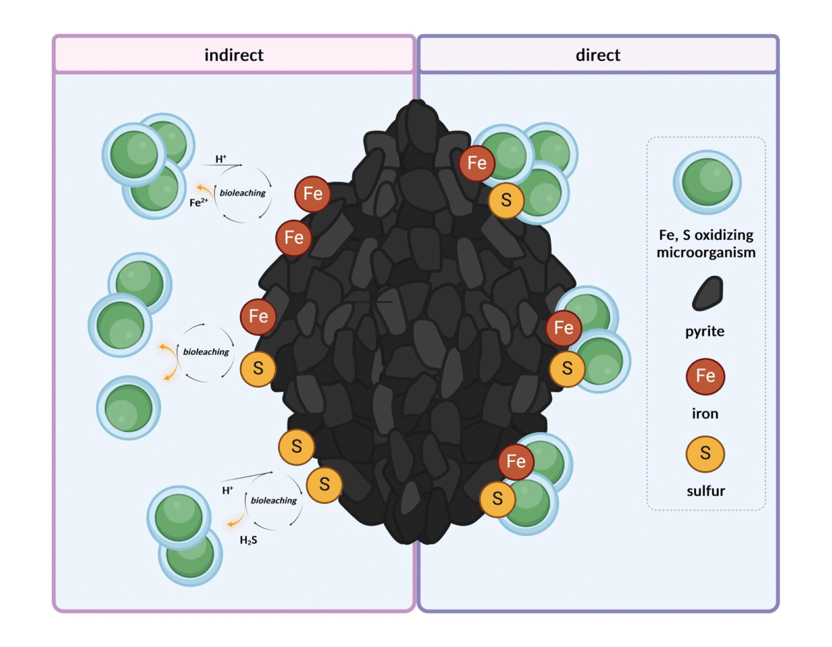
Software is ubiquitous in science, yet overlooked

A recent article in the journal Nature Computational Science, co-authored by fourteen international authors, summarizes the many facets of software in scientific research.

At a time when the scientific world (and beyond) is talking about code, algorithms, and even artificial intelligence, talking about "software" seems to be just another semantic subtlety. Yet there are many facets of software, such as issues of licensing and file formats, that are not part of the definition of code or algorithms. The authors, humanities scholars and practitioners of scientific computing, draw attention to these facets of software that are neglected by researchers, research organizations and funding agencies alike: engineering, governance, licensing, circulation, infrastructure, embedded theory, and users.

Computational chemistry plays an important role in this analysis, because it was one of the first disciplines in which the tension between the industrial and academic approaches to quality assurance became evident. For some, software is reliable if it has been professionally developed according to the best practices of software engineering, while for others, it is the transparency and malleability of Open Source code that guarantees reliability.

See the article:
Hocquet, A., Wieber, F., Gramelsberger, G. et al. Software in science is ubiquitous yet overlooked. Nat Comput Sci (2024). https://doi-org.insb.bib.cnrs.fr/10.1038/s43588-024-00651-2

Protein filaments in the regulation of gene expression

Although every cell in our body contains the same genetic information, cells differ in the way they use it, a process known as “gene expression”. The regulation of gene expression is orchestrated by proteins called transcription factors, which bind to specific sequences within DNA. Transcription factors are traditionally thought to operate mainly as single molecules or dimers.

The article by Mance et al. reveals that several transcription factors of the family known as ZBTB, present in humans and other animals, have the capacity to form non-covalent filamentous structures composed of numerous identical copies of proteins arranged in a chain. At the molecular level, such structures could offer significant advantages for binding to DNA, which is itself an elongated molecule containing numerous repeated sequences. A few examples of filament-forming transcription factors had already been reported, but this study extends the concept to a large family of this protein with important functions. The study - which combines structural, biophysical and functional analysis carried out in vitro and in cells - was carried out by the "Post-translational modifications and DNA repair" team at the CBM and their collaborators in Orléans, Rennes and Marseille, including the "Functional mass spectrometry of molecular assemblies" team also at the CBM.

The findings from this research, together with a complementary study by groups of Benjamin Ebert and Eric Fischer from Dana-Farber Cancer Institute at Harvard (published back-to-back in the same issue of Molecular Cell), challenge the traditional view of transcription factor functionality.

In cells, ZBTB proteins are regulated through a process called SUMOylation, where a small tag called SUMO is added to them, changing how they function. Studies on ZBTB proteins undertaken in Orléans, during which the filamentous structures were discovered, are part of the "SUMOwriteNread" project funded by the European Union (ERC grant no 101078837). Researchers are currently investigating the interplay between the ability to form filaments and SUMO tagging to understand the complex reality of gene expression regulation.

This research was reported by CNRS Chimie on its website.

Dynamic BTB-domain filaments promote clustering of ZBTB proteins.
Lucija Mance, Nicolas Bigot, Edison Zhamungui Sánchez, Franck Coste, Natalia Martín-González, Siham Zentout, Marin Biliškov, Zofia Pukało, Aanchal Mishra, Catherine Chapuis, Ana-Andreea Arteni, Axelle Lateur, Stéphane Goffinont, Virginie Gaudon, Ibtissam Talhaoui, Ignacio Casuso, Martine Beaufour, Norbert Garnier, Franck Artzner, Martine Cadene, Sébastien Huet, Bertrand Castaing & Marcin Józef  Suskiewicz
Molecular Cell 2024
https://doi.org/10.1016/j.molcel.2024.05.029

Dual agents for non-invasive imaging of living organisms

Researchers in the "Lanthanide Luminescent Compounds, Spectroscopy and Optical Bioimaging" team have designed a molecular probe that can image living organisms using both near-infrared (NIR) luminescence and photoacoustic (PA) signal detection. These two complementary imaging techniques make it possible to monitor biological events precisely, in real time and non-invasively.

Find out more

A Dual-Mode Near-Infrared Optical and Photoacoustic Imaging Agent Based on a Low Energy Absorbing Ytterbium Complex
Anton Kovalenko, Svetlana V. Eliseeva, Guillaume Collet, Saïda El Abdellaoui, Sharuja Natkunarajah, Stéphanie Lerondel, Laure Guénée, Céline Besnard & Stéphane Petoud
JACS 2024
https://doi.org/10.1021/jacs.4c03406

Breast cancer: towards early diagnosis by imaging

In vivo imaging of metastatic breast cancer tumors at very early stages is about to become possible. A team of chemists and biologists from the Center for Molecular Biophysics (CNRS) has indeed developed a new magnetic resonance imaging (MRI) probe which has a selective affinity for an emerging biomarker of metastatic breast cancer: Netrin- 1. Find out more on the Cnrs Chimie website.

See more and Cnrs Chmie website.

Reference
Peptide-Conjugated MRI Probe Targeted to Netrin-1, a Novel Metastatic Breast Cancer Biomarker
Clémentine Moreau, Tea Lukačević, Agnès Pallier, Julien Sobilo, Samia Aci-Sèche, Norbert Garnier, Sandra Même, Éva Tóth & Sara Lacerda
Bioconjugate Chemistry 2024
https://doi.org/10.1021/acs.bioconjchem.3c00558

Watching enzymes work in vivo with rare-earth-based molecular probes

Scientists at the CNRS Centre de Biophysique Moléculaire (CBM) in Orléans and the Institut de Chimie des Substances Naturelles (CNRS/Université Paris-Saclay) have designed luminescent probes. They are based on complexes of lanthanides (Ln), a series of rare-earth metals whose trivalent Ln3+ ions are luminescent. The special feature of these probes is that the activity of certain enzymes can modify their luminescence in the near infrared, as well as the signal observed on MRI. These probes make it possible to track the catalytic activity of an enzyme with a single molecule using several complementary imaging techniques: MRI and near-infrared optics. Essential for unambiguous detection of a biological phenomenon, this dual imaging with a single molecule avoids biases due to the use of chemically different imaging agents for each imaging technique.

This study, published in the journal Angewandte Chemie International Edition, paves the way for new non-invasive diagnostic strategies.

This work has been reported on the Cnrs Chimie website

Article reference
Lanthanide-Based Probes for Imaging Detection of Enzyme Activities by NIR Luminescence, T1- and ParaCEST MRI
Rémy Jouclas, Sophie Laine, Svetlana V. Eliseeva, Jérémie Mandel, Frédéric Szeremeta, Pascal Retailleau, Jiefang He, Jean-Francois Gallard, Agnès Pallier, Célia S. Bonnet, Stéphane Petoud, Philippe Durand & Éva Tóth
Angew. Chem. Int. Ed. 2024
https://onlinelibrary.wiley.com/doi/10.1002/anie.202317728

The study of terrestrial fossils in ancient rocks: a crucial approach to identify potential signs of life on Mars

The NASA Perseverance rover is actively exploring Jezero Crater, analyzing igneous and sedimentary rocks from the crater floor and delta deposits. The rock samples that will be returned by the Mars Sample Return (MSR) mission in the 2030s will be subjected to detailed laboratory studies.

Some samples may contain traces of ancient Martian life, which are challenging to detect due to their morphological simplicity and subtle geochemical expressions. Using volcanic sediments from Kitty’s Gap Chert (Pilbara, Australia) of 3.45 billion years as analogues, researchers detail the steps needed to demonstrate their syngenicity and biogenicity. Various analytical methods, including optical and electron microscopy, Raman spectroscopy, X-ray fluorescence spectroscopy, and mass spectrometry, have been employed at different scales. Sedimentological, petrological, mineralogical, and geochemical analyses document a coastal environment of deposition, consistent with the development of microbial life. Morphological, elemental, and molecular analyses of carbonaceous matter associated with potential fossil remnants reveal enrichment in bioessential trace metals (V, Cr, Fe, Co, etc.) and colocalized aromatic and aliphatic molecules of biological origin. This study illustrates the analytical protocol necessary to optimize the detection of fossil traces of life in Martian rocks.

This work is reported on the CNRS Chimie website

Reference
Multi-Technique Characterization of 3.45 Ga Microfossils on Earth: A Key Approach to Detect Possible Traces of Life in Returned Samples from Mars
Laura Clodoré, Frédéric Foucher, Keyron Hickman-Lewis, Stéphanie Sorieul, Jean Jouve, Matthieu Réfrégiers, Guillaume Collet, Stéphane Petoud, Bernard Gratuze, Frances Westall
Astrobiology 2024
http://doi.org/10.1089/ast.2023.0089

Enzymatic detection in near infrared optical imaging and MRI with a single ligand complexed to different lanthanide ions

The imaging visualization of active enzymes is of primary importance in biology.

In a collaborative effort between the Centre of Molecular Biophysics (CBM) and the Institute of Chemistry of Natural Substances (ICSN) in Gif sur Yvette, CBM researchers have designed Ln3+ complexes that provide enzyme-mediated changes in NIR luminescence, as well as in Chemical Exchange Saturation Transfer (CEST) and classical T1-weighted MRI, depending on the Ln3+ used. They have demonstrated the successful monitoring of b-galactosidase activity over time in NIR luminescence and CEST MR imaging in phantoms containing the Yb-complex, and in T1 MRI when using the Gd-analogue. A further great advantage of their probe design is its high versatility, as there are a large number of enzymatically cleavable groups that could be attached to the same core, thus creating probes for other important enzyme targets.

Reference : Rémy Jouclas, Sophie Laine, Svetlana V. Eliseeva, Jérémie Mandel, Frédéric Szeremeta, Pascal Retailleau, Jiefang He, Jean-Francois Gallard, Agnès Pallier, Célia S. Bonnet, Stéphane Petoud, Philippe Durand, Éva Tóth
Lanthanide-Based Probes for Imaging Detection of Enzyme Activities by NIR Luminescence, T1- and ParaCEST MRI
Angew. Chem. Int. Ed. 2024, https://onlinelibrary.wiley.com/doi/10.1002/anie.202317728